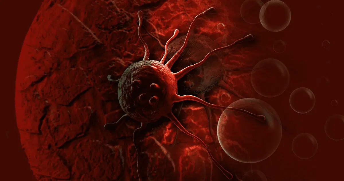
Somewhere in the world, right now, there is a cancer that is thousands of years old.
It isn't a patient's . It's a lineage. It began in one animal long ago and has been passing from dog to dog ever since, outliving every host it has ever lived in. It's one of the oldest continuously living cell lines on Earth, and it's a cancer.
That one fact tells you something important: cancer is not just "a disease." It's an evolutionary process, a rogue cell that learns to survive, hide, spread and, in some strange cases, travel between bodies. In this post we'll follow that story from both sides: how cancer cells cheat, and the creative ways scientists are fighting back. Some of it is physics. Some of it is chemistry. Some of it is borrowed from a shrimp.
Let's start with the basics.
Cancer isn't one disease, it's a playbook
There are more than 200 types of cancer, but they tend to cheat in the same handful of ways. Biologists call these the hallmarks of cancer. A cell doesn't get them all at once. It collects them one by one, like a character leveling up in a game.
- It never stops pressing "grow." Growth-factor receptors like EGFR and HER2, or signaling chains such as RAS-RAF-MEK-ERK and PI3K-AKT-mTOR, get stuck in the "on" position.
- It disables the brakes. like p53 (often called the "guardian of the ") and Rb are supposed to stop damaged cells from dividing. Cancer breaks them.
- It refuses to die. Damaged cells normally self-destruct through apoptosis. Cancer cells dial down pro-death proteins (BAX, BAK) and turn up survival proteins like BCL-2.
- It becomes immortal. Normal cells can divide only so many times because their chromosome ends (telomeres) shorten. Many tumors switch telomerase back on to reset the counter.
- It builds its own supply line. Tumors release VEGF to grow new blood vessels that deliver oxygen and food.
- It learns to travel. Cells lose their "stickiness" (E-cadherin) and release (MMPs) that dissolve the surrounding tissue, so they can escape into the blood and settle in other organs.
Keep this list in mind. Every treatment in this post is an attempt to exploit one of these tricks, or to get around it.
Part 1: The old weapon, and why it's so blunt
Chemotherapy
For decades, the main answer was . The logic is simple: cancer cells divide quickly, so attack anything that divides quickly.
- Alkylating agents (cyclophosphamide, cisplatin) glue strands together, so the cell can't copy its genome and p53-driven self-destruction kicks in.
- Antimetabolites (5-fluorouracil, methotrexate) block enzymes that make the raw materials for DNA, such as thymidylate synthase and DHFR.
- Microtubule drugs (paclitaxel, vincristine) jam the cell's internal scaffolding, so chromosomes can't be pulled apart and cells stall in mitosis.
It works, but it can't tell a cancer cell from any other cell that's busy dividing. That's why side effects follow a pattern:
| Tissue that divides fast | What happens |
|---|---|
| Bone marrow | Low white cells (infection risk), low red cells (exhaustion), low platelets (bleeding) |
| Gut lining | Mouth sores, nausea, vomiting, poor absorption |
| Hair follicles | Hair loss |
| Reproductive cells | Possible temporary or permanent infertility |
So the question researchers asked next was: what if we could make the poison show up only where the cancer is?
The answer: antibody-drug conjugates (ADCs)
Think of an ADC as a guided missile with three parts:
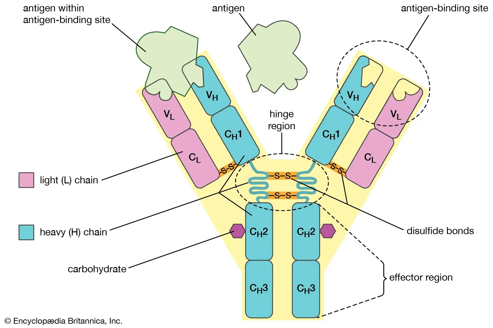
- The guidance system, a (usually IgG1) that recognizes a protein found in large amounts on tumor cells, such as HER2, CD30 or TROP2.
- The coupling, a chemical linker that stays intact in the blood but breaks open once inside the target cell.
- The warhead, a toxin so powerful (often hundreds to thousands of times stronger than ordinary chemo drugs) that it would be unsafe to give on its own. Common ones include auristatins (MMAE), maytansinoids (DM1) and topoisomerase I inhibitors (DXd).
Here's the sequence of events:
- The finds and binds its target on the cancer cell.
- The cell swallows the whole complex (receptor-mediated endocytosis), packing it into a bubble called an endosome.
- The bubble merges with a lysosome, the cell's recycling center. Enzymes like cathepsin B (or the acidic environment) cut the linker.
- The warhead is released and goes straight for its target, either tubulin or DNA.
- The cell dies.
The bonus feature is the bystander effect. Some payloads (MMAE, DXd) are fat-soluble enough to leak out of the dying cell and kill neighboring tumor cells, even ones that lack the target protein. Tumors are rarely uniform, so a drug that can reach "the neighbors" is a huge advantage. Real drugs built on this idea include trastuzumab deruxtecan (HER2), brentuximab vedotin (CD30, used in Hodgkin lymphoma) and sacituzumab govitecan (TROP2).
Part 2: Finding cancer before it announces itself
Treatment is only half the story. Catching cancer early often decides who gets cured.
Blood tests that read tumor DNA
Every cell in your body sheds tiny pieces of DNA into your blood as it dies. These fragments, called cell-free DNA (cfDNA), are usually 140–170 base pairs long, roughly the length of DNA wrapped around one nucleosome. A small share comes from tumors, and that share is called ctDNA.
In early-stage cancer, ctDNA can be under 0.01% of all cfDNA. That's one needle in ten thousand haystacks, which is why detecting it takes extremely sensitive methods like droplet digital PCR and deep sequencing.
Modern "multi-cancer early detection" tests don't rely only on , since not every early tumor carries a known one. They read two more subtle clues:
- Methylation patterns. Tumor DNA carries a distinctive pattern of chemical tags (5-methylcytosine): widespread loss of methylation, plus gain at the promoters of tumor-suppressor . Reading these patterns across millions of sites can reveal both that cancer is present and where it probably started.
- Fragmentomics. Tumor DNA is chopped up differently from healthy DNA, producing unusual fragment lengths and characteristic "end motifs."
The big catch: CHIP. As we age, blood collect harmless mutations in genes like DNMT3A, TET2 and ASXL1, a condition called clonal hematopoiesis of indeterminate potential. Those blood cells shed DNA that can mimic a tumor signal and trigger false alarms. It's a good example of why a test that sounds amazing in principle still needs a lot of careful validation.
PET scans: photographing a tumor's appetite
CT and MRI show what your anatomy looks like. A PET scan shows what your cells are doing, and it does so by exploiting a quirk of cancer metabolism known as the Warburg effect: many tumors burn glucose at a high rate even when plenty of oxygen is available.
The trick has four steps:
- You're injected with FDG, a glucose lookalike tagged with radioactive fluorine-18.
- Hungry tumor cells pull it in through glucose transporters (GLUT1, GLUT3).
- The enzyme hexokinase tags it with a phosphate group, but FDG is missing the chemical feature needed for the next step of glycolysis, so it gets stuck inside the cell.
- Fluorine-18 decays by emitting a positron. When that positron meets an electron, the two annihilate, producing two 511 keV gamma rays that fly off in exactly opposite directions. Detectors ring the patient, and when two of them fire at the same moment, the computer knows the event happened somewhere on the line between them. Millions of these lines build up a 3D map of metabolic activity.
Two practical details: fluorine-18 has a half-life of only about 110 minutes, so FDG has to be made close to where it's used. And some healthy organs, like the brain, also use lots of glucose, so radiologists need to interpret the picture carefully.
Surgery with mantis shrimp vision
During a cancer operation, the surgeon's hardest job is often knowing where the tumor ends. Tumor edges can look much like normal tissue.
Enter the mantis shrimp, an animal with some of the most complex eyes known. Among other things, it can sense the polarization of light, meaning the direction in which light waves oscillate. This turns out to be useful in medicine, because tissue structure changes how light polarizes. Healthy tissue has neatly aligned collagen fibers that reflect light in an orderly manner. Invasive tumors remodel and disorganize that collagen, and it scrambles the polarization.
Engineers have built cameras with microscopic polarizing filters placed over the pixels of a standard CMOS chip. They capture the polarization data (described by Stokes parameters) and convert it into a live color overlay. The goal is to let surgeons see leftover tumor at the margins and remove it, while sparing the healthy tissue around it.
Part 3: Killing tumors with physics
Some tumors can't be removed with surgery, such as those in the liver, kidney, lung or bone, or in patients too frail for an operation. For these, doctors can destroy the tumor in place.
Microwave ablation (MWA) uses a thin antenna, guided by ultrasound or CT, that sits inside the tumor and emits microwaves at 915 MHz or 2.45 GHz. Water molecules are tiny electric dipoles. When the field flips back and forth billions of times per second, they rotate with it, and the friction heats the tissue to somewhere between 60 and 100 °C within seconds. Proteins denature, membranes fall apart, and the tissue dies by coagulation necrosis.
The older technique, radiofrequency ablation (RFA), works differently, by passing an electric current through tissue. The comparison shows why microwaves are attractive:
| RFA | MWA | |
|---|---|---|
| Frequency | 450–500 kHz | 915 MHz / 2.45 GHz |
| How energy travels | Electrical current | Electromagnetic waves |
| Effect of charred tissue | Charring blocks the current and the process stalls | Waves keep spreading through dry, charred tissue |
| Heat sink near big vessels | Flowing blood carries heat away, so tumor cells near vessels can survive | High, fast heating overcomes much of the cooling |
| Ablation zone | Smaller and slower | Larger and faster |
Part 4: Cancer in the wild (it can be contagious)
In humans, cancer is not infectious. In a few animals, it is, and these cases teach us a lot about how tumors dodge the .
The dog tumor that outlived its first dog
Canine transmissible venereal tumor (CTVT) spreads between dogs through mating. Genetic analysis shows that every CTVT tumor descends from a single animal that lived thousands of years ago. It survives in new hosts, who are genetically different from the original, by turning down its MHC molecules, the flags that tell the immune system "this cell is foreign." Without those flags, killer T cells don't recognize it.
The Tasmanian devil's face problem
Tasmanian devils bite each other in the face during fights and mating, and a cancer called devil facial tumor disease (DFTD) has spread through the population this way. Remarkably, there are two separate lineages, DFTD1 and DFTD2. Like CTVT, the tumors hide from the immune system by silencing genes (such as TAP1 and β2-microglobulin) that are needed to display MHC class I on the cell surface. One research direction is to reverse that epigenetic silencing so immune cells can see the tumor again.
Mr. Frosty and the gecko with tumors
In 2015, a leopard gecko with a gorgeous lemon-yellow and white pattern became the founder of a popular color morph, the "Lemon Frost." Then breeders noticed something troubling: the majority of these geckos developed skin tumors called iridophoromas. Those are tumors of iridophores, pigment cells containing light-reflecting guanine crystals.
A genetic study published in 2021 traced the problem to a mutation in the gene SPINT1, which helps control tissue-remodeling signals including the activation of hepatocyte growth factor. SPINT1 is also altered in some human melanomas, so an unplanned breeding experiment ended up giving scientists a natural animal model of pigment-cell cancer.
Part 5: How tumors adapt to hostile places
Breast cancer in the brain
When breast cancers (especially HER2-positive and triple-negative types) spread to the brain, they face two huge barriers.
First, the blood-brain barrier. Brain capillaries are lined with tightly joined cells (held together by proteins such as claudins and occludins). Cancer cells have to stick to the vessel wall and then release enzymes like cathepsin S and matrix metalloproteinases to pry the barrier open.
Second, a hostile neighborhood. Once inside, they meet reactive astrocytes that attack invaders. Some surviving breast cancer cells adapt in a strange way: they take up GABA, the brain's main inhibitory neurotransmitter, and break it down using the enzyme GABA transaminase. The leftover carbon enters their energy cycle as succinate. In other words, they learn to eat a neurotransmitter to survive in an environment where normal food is scarce.
The iron addiction
Cancer cells need a lot of iron, for building DNA (ribonucleotide reductase relies on it) and for powering . That need makes them vulnerable.
Iron can also drive a dangerous chemical reaction, the Fenton reaction:
Fe²⁺ + H₂O₂ → Fe³⁺ + •OH + OH⁻
That produces a hydroxyl radical, which tears into the fatty acids in cell membranes and starts a chain reaction of lipid peroxidation. Cells defend themselves with an enzyme called GPX4, which uses glutathione to neutralize those damaged lipids.
Ferroptosis, a form of cell death described in 2012, happens when that defense fails and iron-driven lipid damage runs away. Researchers can trigger it experimentally with compounds like erastin (blocks cystine import through System xc⁻, which lowers glutathione) and RSL3 (inhibits GPX4 directly). It's particularly exciting because some drug-resistant cancer cells seem to be unusually sensitive to ferroptosis. It's still early-stage science, though, and the challenge is doing this selectively without harming healthy tissue.
Part 6: Rethinking what "type of cancer" means
Traditionally, cancer was classified by where it started: lung, breast, colon. Sequencing has changed that. Two tumors from different organs can share the same driver mutation, and two tumors from the same organ can be completely different.
That led to tissue-agnostic therapies, drugs approved based on a tumor's molecular feature rather than its location:
- Pembrolizumab (Keytruda) was approved for solid tumors with MSI-high or mismatch repair deficiency (dMMR), regardless of organ. This was the first approval of its kind, in 2017.
- Larotrectinib and entrectinib target tumors with NTRK gene fusions.
To test such drugs, trial designs had to change too:
- Basket trials take one drug and test it on many cancer types that share one mutation (for example, BRAF V600E in melanoma, colorectal and lung cancers).
- Umbrella trials do the reverse: they take a single cancer type and assign patients to different drugs depending on their tumor's genetic profile.
Part 7: Prevention, and the diet soda panic
What actually moves the needle
Roughly 40% of cancers worldwide are linked to causes we can change. The big ones:
- Tobacco. Smoke contains more than 70 known carcinogens, and it leaves characteristic mutations in genes like TP53 and KRAS.
- Alcohol. The body converts ethanol into acetaldehyde, which damages DNA and interferes with its repair.
- UV radiation. UVB causes abnormal DNA bonds (cyclobutane pyrimidine dimers), leaving the signature mutations seen in skin cancers.
- Excess body fat. It's associated with chronic low-grade inflammation, higher insulin and IGF-1, and more estrogen production in fat tissue, all of which help drive breast and endometrial cancers.
- Infections. Around one in eight cancers worldwide are linked to infections. High-risk HPV produces E6 and E7 proteins that disable p53 and Rb, and the HPV vaccine (Gardasil 9) provides strong protection. Hepatitis B infection can lead to liver cancer, and the HBV helps prevent it.
Aspartame: a lesson in reading headlines
In 2023, news outlets reported that the WHO had classified aspartame as "possibly carcinogenic." Panic followed. But two different WHO-linked groups had weighed in, and they were answering two different questions:
- IARC asks a hazard question: can this thing cause cancer under any conditions? It put aspartame in Group 2B ("possibly carcinogenic").
- JECFA asks a risk question: is it likely to harm you at normal exposure levels? It left the acceptable daily intake unchanged at 40 mg per kg of body weight.
| IARC group | Meaning | Examples |
|---|---|---|
| Group 1 | Carcinogenic to humans | Tobacco smoke, asbestos, UV radiation, processed meat |
| Group 2A | Probably carcinogenic | Red meat, very hot beverages, night-shift work |
| Group 2B | Possibly carcinogenic | Aspartame, aloe vera extract, some pickled vegetables |
| Group 3 | Not classifiable | Many everyday exposures with insufficient evidence |
The math: for a 70 kg adult, the limit is about 2,800 mg per day. A can of diet soda contains roughly 200–300 mg, so you'd have to drink about 9–14 cans a day to exceed it. (Separately, people with the rare condition phenylketonuria need to avoid aspartame for a different reason, because it contains phenylalanine.) And remember that classification groups describe the strength of evidence, not how dangerous something is. Processed meat and tobacco smoke are in the same group, but their risks are not remotely similar.
Part 8: The body's early warning system
Many people say, after a cancer diagnosis, that they felt "off" for months, tired in a way they couldn't explain. Could there be a real basis for that?
Possibly, through interoception, the nervous system's sense of the body's internal state. Growing tumors disturb local metabolism and trigger inflammation, releasing signals like IL-1β, IL-6 and TNF-α. The body can pick these up in two ways:
- A neural route. Sensory fibers of the vagus nerve carry inflammation signals to the brainstem (nucleus tractus solitarius).
- A chemical route. Cytokines in the blood reach brain regions that lack a tight blood-brain barrier, such as the area postrema, and stimulate production of prostaglandin E2.
Both routes feed into the hypothalamus and produce what's called sickness behavior: deep fatigue, changes in sleep, loss of appetite, reduced pleasure, and shifts in body temperature. It's not a supernatural premonition. It's the immune system and brain communicating, and it's a good argument for not ignoring persistent changes that have no obvious explanation.
What it all adds up to
Step back and look at what we've covered:
- Toxins delivered like guided missiles
- DNA fragments in a blood tube
- Antimatter annihilation to image a tumor's sugar habit
- A camera copied from a crustacean
- Microwaves heating water inside a tumor
- Cancers that spread like infections
- Cells that eat brain chemicals
- A cell-death program fueled by iron
It's tempting to think of cancer research as a single battle. It's really a set of many small puzzles, solved by people from physics, chemistry, genomics, evolution, engineering and neuroscience. Not everything here is a finished clinical tool. Liquid-biopsy screening is still being validated, ferroptosis drugs are largely experimental, and polarization cameras are still emerging. But the direction of travel is encouraging: earlier detection, more precise targeting, and gentler treatment.
And the most hopeful thing may be the simplest: the more we understand how cancer cheats, the better we get at beating it.
In the literature
PubMed and arXiv
- PubMed2020The Frequency of Ras Mutations in Cancer.Prior IA, Hood FE, Hartley JL · Cancer researchRead the paper
- PubMed2024Targeting MTHFD2 to Exploit Cancer-Specific Metabolism and the DNA Damage Response.Ramos L, Henriksson M, Helleday T · Cancer researchRead the paper
- PubMed2014Projecting cancer incidence and deaths to 2030: the unexpected burden of thyroid, liver, and pancreas cancers in the United States.Rahib L, Smith BD, Aizenberg R · Cancer researchRead the paper
- PubMed2025SMAD4 Deficiency Promotes Pancreatic Cancer Progression and Confers Susceptibility to TGFβ Inhibition.Murimwa GZ, Williams NE, Alzhanova D · Cancer researchRead the paper
Matched automatically on this article’s subjects. Relevance varies — these are suggestions for further reading, not sources for the piece above.
Topics
Sign in to save articles and join the discussion.