Restriction Endonucleases:
1. Definition and Basic Concept
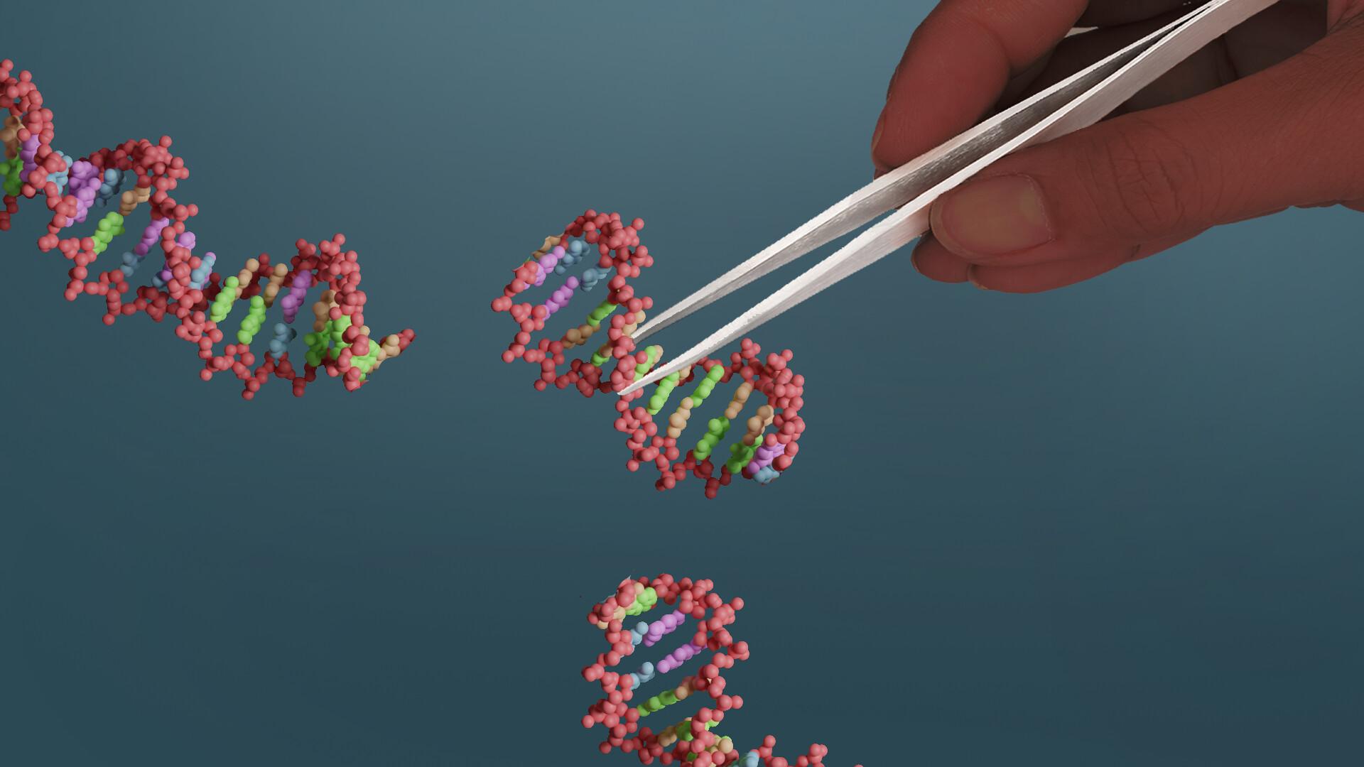
Restriction endonucleases are specialized that cleave double-stranded at specific recognition sequences. These molecular scissors play a crucial role in various cellular processes and have become indispensable tools in molecular biology and biotechnology.
- Origin: Primarily found in bacteria and archaea
- Function in nature: Act as a defense mechanism against foreign DNA, particularly from bacteriophages
- Discovery: First identified in the 1970s by Werner Arber, Hamilton O. Smith, and Daniel Nathans
2. Historical Context
- 1950s: Salvador Luria and Giuseppe Bertani observe phenomenon of "host-controlled restriction"
- 1960s: Werner Arber proposes the existence of restriction enzymes
- 1970: Hamilton O. Smith isolates and characterizes the first type II restriction enzyme (HindII from Haemophilus influenzae)
- 1971: Daniel Nathans demonstrates the use of restriction enzymes to analyze viral DNA
- 1978: Arber, Smith, and Nathans awarded the Nobel Prize in Physiology or Medicine
3. Types of Restriction Endonucleases
Restriction endonucleases are classified into four main types based on their subunit composition, cofactor requirements, recognition sequences, and cleavage positions:
Type I
- Structure: Complex with three subunits - R (restriction), M (modification), and S (sequence specificity)
- Cofactors: Require Mg2+, ATP, and S-adenosyl methionine (SAM)
- Recognition: Asymmetric, bipartite sequences
- Cleavage: At random sites, often far (up to thousands of base pairs) from recognition sequence
- Mechanism: ATP-driven DNA translocation before cleavage
- Examples: EcoKI, EcoBI
Type II
Type II restriction endonucleases are the most widely used in molecular biology and genetic engineering due to their simplicity and predictability. They have several key characteristics that make them particularly useful:
Structure and Composition
- Usually function as homodimers (two identical subunits)
- Each subunit typically contains a catalytic domain and a DNA-binding domain
- Some Type II enzymes have a more complex quaternary structure
Cofactor Requirements
- Require Mg2+ as a cofactor for catalytic activity
- Do not require ATP for DNA cleavage
Recognition Sequences
- Recognize specific DNA sequences, typically 4-8 base pairs in length
- Most recognition sequences are palindromic (read the same in both directions on complementary strands)
- Example: EcoRI recognition sequence is 5'-GAATTC-3' 3'-CTTAAG-5'
- Some Type II enzymes recognize non-palindromic sequences
Cleavage Characteristics
- Cut DNA at precise positions within or near their recognition sites
- Can produce either blunt ends or sticky (cohesive) ends
- Blunt ends: Cut straight across both strands (e.g., SmaI cuts 5'-CCC^GGG-3')
- Sticky ends: Staggered cuts leaving single-stranded overhangs (e.g., EcoRI cuts 5'-G^AATTC-3') 3'-CTTAA^G-5'
Mechanism of Action
- DNA Binding: The enzyme scans the DNA and binds to its specific recognition sequence.
- Conformational Change: Binding induces a conformational change in the enzyme, activating the catalytic site.
- Phosphodiester Bond Hydrolysis: The enzyme cleaves the phosphodiester bonds in the DNA backbone.
- Product Release: The enzyme releases the cleaved DNA fragments.
Subtypes of Type II Restriction Endonucleases
- Type IIP: Classic palindromic-recognizing enzymes (e.g., EcoRI, BamHI)
- Type IIS: Cut outside their asymmetric recognition sequences (e.g., FokI)
- Type IIE: Require two recognition sites for cleavage (e.g., EcoRII)
- Type IIF: Cleave both recognition sites concertedly (e.g., SfiI)
- Type IIB: Cleave on both sides of the recognition sequence (e.g., BcgI)
- Type IIT: Composed of two different subunits (e.g., Bpu10I)
- Type IIG: Contain both cleavage and modification domains in a single polypeptide (e.g., Eco57I)
Type III
- Structure: Two subunits - Res (restriction) and Mod (modification)
- Cofactors: Require Mg2+ and ATP
- Recognition: Asymmetric sequences
- Cleavage: 25-27 base pairs downstream of recognition site
- Mechanism: ATP-dependent DNA translocation before cleavage
- Examples: EcoP1I, EcoP15I
Type IV
- Structure: Single subunit
- Recognition: Modified (typically methylated) DNA
- Cleavage: At variable distances from recognition site
- Examples: McrBC, Mrr
Regulation in Bacterial Cells
- Often paired with methyltransferases in restriction-modification systems
- Methyltransferases protect host DNA by adding methyl groups to the recognition sequences
- This system allows bacteria to distinguish between self and non-self DNA
Applications in Molecular Biology
- DNA Cloning: Creating recombinant DNA molecules
- Genetic Engineering: Inserting, deleting, or modifying
- Mapping: Creating physical maps of genomes
- Southern Blotting: Detecting specific DNA sequences
- RFLP Analysis: Studying genetic variations between individuals
- DNA Fingerprinting: Used in forensics and paternity testing
- Construction of DNA Libraries: Creating collections of DNA fragments
Regulation and Control in Bacterial Cells
- Often part of restriction-modification systems
- Paired with methyltransferases to protect host DNA from cleavage
Commercial Production and Use
- Produced through recombinant DNA technology in E. coli
- Available commercially for research and biotechnology applications
- Stored and used under specific buffer and temperature conditions
Challenges and Limitations
- Star activity: Non-specific cutting under suboptimal conditions
- Methylation sensitivity: Some enzymes are blocked by DNA methylation
- Incomplete digestion: Can occur due to various factors (enzyme quality, reaction conditions, etc.)
Recent Advances and Future Prospects
- Engineering of novel restriction enzymes with custom recognition sites
- Use in genome editing technologies (e.g., as part of -Cas systems)
- Potential applications in personalized medicine and diagnostics
DNA-Modifying Enzymes
1. DNA Ligases
DNA ligases are essential enzymes that catalyze the formation of phosphodiester bonds between the 3'-hydroxyl end of one DNA strand and the 5'-phosphate end of another. They play a crucial role in DNA replication, repair, and recombination.
Structure and Composition
- DNA ligases are typically monomeric
- Eukaryotic ligases are larger (90-130 kDa) than prokaryotic ligases (70-80 kDa)
- Contain multiple domains:
- Nucleotidyltransferase (NTase) domain: Catalytic core
- Oligonucleotide-binding (OB) fold domain: DNA binding
- DNA-binding domain (DBD): Enhances specificity and efficiency
- BRCT domain (in eukaryotes): Protein-protein interactions
Mechanism of Action
- Activation: ATP (eukaryotes) or NAD+ (prokaryotes) binds to the enzyme
- Nucleotidyl transfer: AMP is transferred to a lysine residue in the active site
- DNA binding: Enzyme binds to nicked DNA
- Adenylyl transfer: AMP is transferred from the enzyme to the 5'-phosphate of DNA
- Phosphodiester bond formation: 3'-OH attacks the adenylated 5'-phosphate, forming a new bond
- Release: AMP is released, and the enzyme dissociates from the sealed DNA
Types of DNA Ligases
- ATP-dependent ligases: Found in eukaryotes, , and some bacteria
- Examples: T4 DNA ligase, human DNA ligase I, II, III, and IV
- NAD+-dependent ligases: Primarily found in bacteria
- Example: E. coli DNA ligase
Characteristics
- Require a divalent metal ion (usually Mg2+) as a cofactor
- Optimal temperature varies (37°C for mammalian ligases, 16°C for T4 DNA ligase)
- pH optimum typically between 7.5-8.0
- Can join both blunt and cohesive ends, but cohesive ends are ligated more efficiently
Applications in Molecular Biology
- Molecular cloning: Joining insert DNA to DNA
- Next-generation sequencing: Library preparation
- DNA repair studies
- Ligase chain reaction (LCR) for DNA amplification and detection
2. DNA Polymerases
DNA polymerases are enzymes responsible for synthesizing DNA molecules by assembling nucleotides. They are crucial for DNA replication, repair, and various molecular biology techniques.
Structure and Composition
- Typically large, multi-subunit enzymes
- Structure often described as resembling a "right hand" with three domains:
- Palm domain: Contains the catalytic site
- Thumb domain: Interacts with the DNA minor groove
- Fingers domain: Interacts with the incoming nucleoside triphosphate
Types of DNA Polymerases
- Replicative polymerases: High processivity and fidelity
- Examples: Pol δ and Pol ε (eukaryotes), Pol III (prokaryotes)
- Repair polymerases: Lower processivity, specialized functions
- Examples: Pol β, Pol λ, Pol μ (base excision repair)
- Translesion synthesis (TLS) polymerases: Can bypass DNA damage
- Examples: Pol η, Pol κ, Pol ι
Mechanism of Action
- Template binding: Enzyme binds to single-stranded DNA template
- Primer binding: Short or DNA primer anneals to template
- Nucleotide selection: Incoming dNTP pairs with template base
- Phosphodiester bond formation: 3'-OH of primer attacks α-phosphate of dNTP
- Translocation: Enzyme moves to the next template base
Characteristics
- 5' to 3' synthesis direction
- Require a primer with a free 3'-OH group
- Most have 3' to 5' exonuclease activity for proofreading
- Some have 5' to 3' exonuclease activity for removing RNA primers
Commonly Used DNA Polymerases in Molecular Biology
-
Taq polymerase (from Thermus aquaticus)
- Thermostable, used in PCR
- Lacks 3' to 5' exonuclease activity
- Error rate: ~1 in 10^4-10^5 nucleotides
-
Pfu polymerase (from Pyrococcus furiosus)
- Higher fidelity than Taq due to 3' to 5' exonuclease activity
- Error rate: ~1 in 10^6 nucleotides
-
Phi29 DNA polymerase
- Highly processive, used in whole genome amplification
- Has strand displacement activity
-
T4 DNA polymerase
- High 3' to 5' exonuclease activity
- Used for creating blunt ends and removing 3' overhangs
Applications in Molecular Biology
- Polymerase Chain Reaction (PCR)
- DNA sequencing (Sanger method)
- Site-directed mutagenesis
- Molecular cloning
- DNA labeling and detection
3. Polynucleotide Kinase
Polynucleotide kinase (PNK) is an enzyme that catalyzes the transfer of a phosphate group from ATP to the 5'-hydroxyl terminus of DNA or RNA. It also has 3'-phosphatase activity.
Structure and Composition
- Monomeric enzyme in most organisms
- Contains three domains:
- N-terminal fused kinase-phosphatase domain
- Kinase domain
- Phosphatase domain
Mechanism of Action
- Kinase activity:
- ATP binds to the enzyme's active site
- 5'-OH of DNA/RNA attacks the γ-phosphate of ATP
- Phosphate is transferred, creating a 5'-phosphate on the nucleic acid
- Phosphatase activity:
- 3'-phosphate is hydrolyzed, leaving a 3'-OH group
Characteristics
- Requires Mg2+ as a cofactor
- Optimal pH: 7.5-8.0
- Can phosphorylate both DNA and RNA
- More efficient with single-stranded substrates, but can act on double-stranded molecules
Applications in Molecular Biology
- 5' end-labeling of DNA/RNA with radioactive phosphate
- Preparing DNA for ligation (adding 5'-phosphate)
- Removing 3'-phosphates from DNA/RNA
- DNA repair studies
4. Alkaline Phosphatase
Alkaline phosphatase (AP) is a hydrolase enzyme that removes phosphate groups from many types of molecules, including nucleotides, proteins, and alkaloids. In molecular biology, it's primarily used to remove 5'-phosphate groups from DNA and RNA.
Structure and Composition
- Homodimeric enzyme
- Each subunit contains:
- Two Zn2+ ions and one Mg2+ ion in the active site
- A serine residue crucial for catalytic activity
Types of Alkaline Phosphatases
- Bacterial APs (e.g., E. coli AP)
- Mammalian APs:
- Tissue-nonspecific AP (TNAP)
- Intestinal AP (IAP)
- Placental AP (PLAP)
- Germ cell AP (GCAP)
- Shrimp AP: Heat-labile, used in molecular biology
Mechanism of Action
- Substrate binding: Phosphorylated molecule binds to the active site
- Nucleophilic attack: Serine residue attacks the phosphate group
- Phosphate transfer: Phosphate is transferred to the enzyme, forming a covalent intermediate
- Hydrolysis: Water molecule hydrolyzes the phospho-enzyme intermediate
- Product release: Dephosphorylated substrate and inorganic phosphate are released
Characteristics
- Optimal pH: 8.0-10.5 (hence "alkaline")
- Requires Zn2+ and Mg2+ as cofactors
- Heat-stable enzymes (bacterial and calf intestinal AP) retain activity after heat-inactivation of other enzymes
- Can be inactivated by heat (65°C for shrimp AP, 85°C for calf intestinal AP)
Applications in Molecular Biology
- Dephosphorylating DNA vectors to prevent self-ligation
- Removing 5'-phosphates from RNA or DNA prior to end-labeling
- Dephosphorylating nucleotides in sequencing reactions
- Used as a reporter enzyme in ELISA and immunohistochemistry
5. Nucleases
Nucleases are enzymes that cleave the phosphodiester bonds between nucleotide subunits of nucleic acids. They play crucial roles in various cellular processes and are widely used in molecular biology techniques.
Classification of Nucleases
-
Based on substrate specificity:
- DNases: Cleave DNA
- RNases: Cleave RNA
- Non-specific nucleases: Can cleave both DNA and RNA
-
Based on cleavage pattern:
- Endonucleases: Cleave within the nucleic acid chain
- Exonucleases: Cleave from the ends of nucleic acid molecules
Structure and Composition
- Vary widely depending on the specific nuclease
- Often contain metal ions (e.g., Mg2+, Zn2+) in their active sites
- May be monomeric, dimeric, or multimeric
Mechanism of Action (General)
- Substrate binding: Nuclease binds to nucleic acid
- Activation of water molecule: Metal ion cofactor activates a water molecule
- Nucleophilic attack: Activated water attacks the phosphodiester bond
- Bond cleavage: Phosphodiester bond is broken
- Product release: Cleaved nucleic acid fragments are released
Characteristics and Examples
DNases
-
DNase I
- Endonuclease that preferentially cleaves double-stranded DNA
- Requires Mg2+ and Ca2+ for activity
- Generates 5'-phosphorylated oligonucleotides
- Applications: Removing contaminating DNA, footprinting assays
-
DNase II
- Endonuclease active in acidic conditions (pH optimum ~4.5-5.0)
- Generates 3'-phosphorylated oligonucleotides
- Role in apoptosis and degradation of exogenous DNA
-
Restriction Endonucleases
- Recognize specific DNA sequences (covered in detail earlier)
RNases
-
RNase A
- Endoribonuclease specific for single-stranded RNA
- Cleaves after pyrimidine residues
- Heat-stable enzyme
- Applications: Removing RNA contamination, RNA structure studies
-
RNase H
- Endonuclease that specifically degrades RNA in RNA-DNA hybrids
- Important for DNA replication and reverse transcription
- Applications: Removing RNA primers in cDNA synthesis
-
RNase III
- Cleaves double-stranded RNA
- Important in RNA processing and gene silencing
- Dicer (a type of RNase III) is crucial in the RNAi pathway
Non-specific Nucleases
-
S1 Nuclease
- Single-strand specific nuclease that can degrade both DNA and RNA
- Requires Zn2+ for activity
- Applications: Mapping single-stranded regions in DNA, removing single-stranded overhangs
-
Mung Bean Nuclease
- Similar to S1 nuclease but more specific for single-stranded DNA
- Applications: Creating blunt ends from cohesive ends
Applications in Molecular Biology
- DNA/RNA isolation and purification
- Mapping of DNA-protein interactions (e.g., DNase I footprinting)
- Generation of defined DNA fragments (restriction endonucleases)
- RNA interference studies
- Removal of nucleic acid contamination
- Structural studies of nucleic acids
6. Reverse Transcriptase
Reverse transcriptase (RT) is an enzyme that synthesizes complementary DNA (cDNA) using an RNA template. Originally discovered in retroviruses, it has become an essential tool in molecular biology.
Structure and Composition
- Typically consists of two subunits:
- Larger subunit (~65-90 kDa): Contains the polymerase and RNase H domains
- Smaller subunit (~50-65 kDa): Enhances processivity and promotes template switching
Domains and Activities
- RNA-dependent DNA polymerase activity
- Synthesizes cDNA using RNA as a template
- DNA-dependent DNA polymerase activity
- Can use single-stranded DNA as a template
- RNase H activity
- Degrades RNA in RNA-DNA hybrids
Mechanism of Action
- Primer binding: RT binds to a primer (often oligo-dT or random hexamers) annealed to RNA template
- cDNA synthesis: RT synthesizes DNA in a 5' to 3' direction, using dNTPs
- Template switching: When RT reaches the 5' end of RNA, it can switch to the newly synthesized cDNA strand
- Second-strand synthesis: RT uses the first cDNA strand as a template to create double-stranded cDNA
Characteristics
- Lacks 3' to 5' exonuclease proofreading activity
- Error rate: ~1 in 10^4-10^5 nucleotides
- Requires Mg2+ as a cofactor
- Optimal temperature varies (37°C for AMV RT, 42°C for M-MuLV RT)
Types of Reverse Transcriptases
-
Avian Myeloblastosis Virus (AMV) RT
- High thermal stability (up to 55°C)
- Efficient for templates with high secondary structure
-
Moloney Murine Leukemia Virus (M-MuLV) RT
- Lower RNase H activity compared to AMV RT
- Better for synthesizing longer cDNAs
-
Engineered RTs
- Engineered for improved thermal stability, processivity, and fidelity
- Examples: SuperScript enzymes, ThermoScript RT
Applications in Molecular Biology
- cDNA synthesis for RT-PCR and RT-qPCR
- Construction of cDNA libraries
- RNA sequencing (RNA-seq)
- Analysis of gene expression
- Detection of RNA viruses
- RACE (Rapid Amplification of cDNA Ends) for determining full-length RNA sequences
Challenges and Considerations
- RNA secondary structure can impede RT activity
- Some RTs may add non-templated nucleotides at the 3' end of cDNA
- The choice of primers (oligo-dT, random hexamers, or gene-specific) affects the resulting cDNA population
- RNase H activity can be both beneficial (allowing for more efficient synthesis of the second DNA strand) and detrimental (potentially degrading the RNA template before complete cDNA synthesis)
In the literature
PubMed and arXiv
- PubMed1994Molecular biology of breast cancer.Lemoine NR · Annals of oncology : official journal of the European Society for Medical OncologyRead the paper
- PubMed1994Bridging the gap between physiology and molecular biology: new approaches to perpetual questions.Norwood VF, Gomez RA · The American journal of physiologyRead the paper
- PubMed2015Molecular cloning and bioinformatic analysis of the Streptococcus agalactiae neuA gene isolated from tilapia.Wang EL, Wang KY, Chen DF · Genetics and molecular research : GMRRead the paper
- PubMed1988Molecular cloning, sequencing, and mapping of the bacteriophage T2 dam gene.Miner Z, Hattman S · Journal of bacteriologyRead the paper
Matched automatically on this article’s subjects. Relevance varies — these are suggestions for further reading, not sources for the piece above.
Sign in to save articles and join the discussion.